Mechanism and subjective history
The MCL (Medial Collateral Ligament) is stressed with a valgus +/- an external rotation force to the knee. A typical mechanism for this is a block tackle, which can include an external rotation element. Landing and change of direction may also be reported as the cause. The superficial MCL fibres are the primary restraint to valgus forces, however if an external rotation element is included, the deep fibres of the MCL may be injured, since these are the primary restraint to tibial external rotation.
The individual will commonly present with focal, medial pain and complain of the knee feeling wobbly, loose or unstable.
Deep fibre tears may initially present as being more painful than superficial tears, likely due the close anatomical connection to the joint capsule and medial meniscus.
A low-grade MCL injury may be tolerated to allow an individual to continue. However, a feeling of soreness and stiffness after will be reported, as opposed to any significant instability symptoms.
Individuals with higher-grade 2+ injuries will be unlikely to continue, reporting an immediate pain and feeling of instability.
Click here to see a famous MCL injury – Cristiano Ronaldos knee injury in the final of Euro 2016. He tried to carry on no doubt hoping for a Grade 1 but was unable to do so and had to be removed shortly after.
Objective examination
Observation
There will unlikely be an effusion in an isolated MCL injury. Higher grades may present with localised medial knee swelling and bruising, which may track down to the medial shin.
Palpation
Should aim to identify the key areas of tenderness at the MCL. The posterior oblique ligament should be palpated which is located posteriorly to the superficial MCL fibres.
ROM
Should be assessed through range and pain response noted.
Stability
MCL valgus stress at 0° and 30° flexion is tested. External tibial rotation should be added to assess the rotational stability of the knee, which is mostly contributed by the deep fibres of the MCL and posterior oblique ligament, particularly closer to full extension. Distal tears of the superficial ligament are likely to present as being more stable given the long attachment, approximately 6cm below the joint line (Kowalczuk et al., 2018). Deep fibre injury may present with less valgus instability, however, should not be disregarded as a less serious injury considering the risk of complications and ongoing symptoms (Kowalczuk et al., 2018).
Grade 1: 0-5mm gapping
Grade 2: 5-10mm gapping
Grade 3: >10mm gapping
Click here to see an excellent demonstration of the Valgus stress testing of the medial structures of the knee.
Other key tests
- All other knee ligament tests should be completed. An ACL injury may have a similar mechanism to that of an MCL. The dial test should be used to assess the rotational stability of the knee.
- Meniscal examination: The medial meniscus may be involved, especially with a rotational element to the mechanism. The lateral meniscus should also be assessed considering the potential compressive forces at the lateral compartment.
Radiology
Key information from MRI report:
- Integrity of superficial and deep MCL fibres. Presence of oedema and any structural damage. Characteristics of fibre damage, including dimensions, along with any loss of tension.
- Medial meniscus, medial joint capsule, meniscotibial and meniscofemoral ligaments.
- Posterior oblique ligament, semi membranosus and popliteal oblique ligament at the posterior medial knee.
- Exclude bony avulsions, associated tibial plateau and distal femur fractures.
Key components of rehabilitation PROTECT, STIFFEN, STRENGTHEN
The MCL is extra-articular and well vascularised
The four stages of healing of haemorrhage, inflammation, repair and remodelling should be maximised with early, diligent bracing, to facilitate the early healing stages with the aim to maximise the restoration of the stability in the ligament.
To brace or not to brace
This is often a contentious area with differing opinions regarding the pros and cons of bracing. In our opinion bracing protects the knee when the patient isn’t thinking about it (turning with a relaxed knee, turning in bed etc) whilst allowing an aggressive strengthening regime within the limitations of the brace settings.
Bracing allows the healing ligament to be gradually stressed to stimulate remodelling. The use of the Alter-G treadmill allows weight bearing status to be progressed earlier and running introduced earlier too.
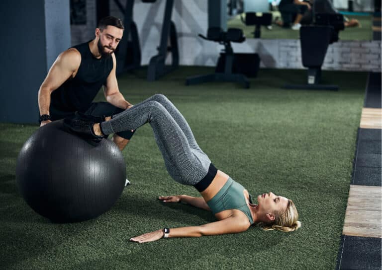
Active stability around the knee is maximised early
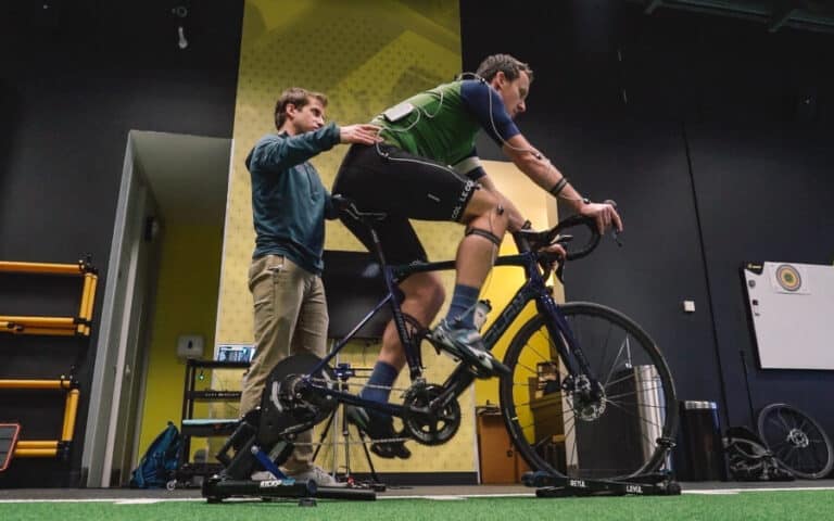
The individual should begin localised strengthening to ensure active stabilisation of the knee is maintained. This can be completed safely using the IKD to complete quadriceps and hamstring strength while still in the brace and complying with weightbearing and range of motion restrictions. This can be incorporated into a global gym programme working within the limitations of the bracing guidelines.
Compex and Blood Flow Restriction training
Muscle stimulation and occlusion training should be incorporated into the rehabilitation program. This will limit muscle atrophy and maximise hypertrophy during the appropriate phase of training.
Delay multi-plane tasks
Change of direction and multi-plane tasks, including kicking progressions, should be delayed until adequate stability in the ligament is achieved with minimal remaining symptoms. This is tested at the point of brace removal, with the laxity grade compared with that post-injury, and the opposite knee.
Although complete stiffness of the ligament will unlikely be reached at this point, minimal pain and a feeling of stability should be reported by the patient. At this point, a grade 1 like presentation should be present, with the assumption that adequate healing has occurred and progressive stress to the ligament will now be advantageous to the remodelling stage.
Delay kicking and block tackling
Although progressive exposure to kicking and block tackling is incorporated at the end stage, maximal testing of this action is left until the final sessions. This allows the healing ligament maximal healing time and increases the chances of a successful mechanical clearance before RTP.
Ligament stability and knee function testing
The knee is likely to be left with increased laxity compared to pre-injury. This becomes the new normal for that individual. As long as there is a firm end feel a small increase in laxity is considered normal and will continue to improve and stiffen as the scar tissue continues to model over the next 6 to 8 months.
Along with passive knee stability the overall function of the knee should also be tested prior to return to full activity and will include
- IKD conc quadriceps 3 x BW and 10% LSI
- IKD ecc hamstrings 2.5 x BW and 10% LSI
- SL max compound strength return to baseline
- Planned and unplanned cutting tasks
- DL and SL CMJ and Drop jump within 10% of baseline
- Horizontal and lateral hops within 10% of baseline
- Peak plantar force 10% LSI
- HHD Ab/Ad 10% LSI
So how long will you be injured??
Medial knee injuries are the most common knee injury in professional football. The make up 4% of all injuries in football and have an occurrence of approx 0.33 per 1000 hours of activity.
In terms of how long the recovery takes it will be impacted by the structures involved, whether it’s isolated to deep, superficial fibres and involvement of other stabilising structures in the medial and posteromedial aspect of the knee.
Typically the time scale given is around 7-10 weeks though a good rule of thumb we apply based on our own experience with these injuries in the last 20 years is 50 days. The majority of these injuries with variation of injection therapies utilised (no injection, PRP injection, Prolotherapy injection) return to full contact activity at around 50 days if a progressive rehabilitation program following the principles of PROTECT-STIFFEN-STRENGTHEN-MEASURE is followed.
The final phase of any rehabilitation program should of course include gradual exposure to the key physical components of the sport or activity the individual is returning to and integration into training and finally match activity as appropriate.